

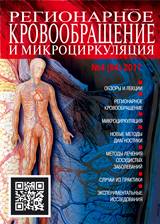
Academic and research Journal «Regional Blood Circulation and Microcirculation» is a multidisciplinary research and practice journal that provides a platform for the publication of research in a broad range of medical fields and specialties, including all branches of cardiovascular research both experimental ones and related to different areas of clinical medicine such as cardiology, cardiovascular surgery, radiology, neurology etc.
To be accepted for publication the submitted material should be earlier unpublished and correspond to the journal's area of expertise: reviews, and results of completed original clinical and experimental studies, lectures, descriptions of clinical cases, book reviews, readers' comments on earlier published papers, letters to the editor, information on scientific events.
The Journal is registered in the Ministry of the Russian Federation for Affairs of the Press, Television and Radio Broadcasting and Mass Communication Media.
The Certificate about the registration of mass media «ПИ № 77-9025» from 21.05.2001.
The Journal is the official organ of the Association for Microcirculation and Regional Hemodynamics that is part of the European Society for Microcirculation.
The editorial board and editorial review board of the Journal consist of 30 scientists from top-ranked Russian universities and research centers, including 5 academicians and 6 corresponding members of the RAS. The international editorial review board includes 5 experts from Great Britain, Norway, Germany, Portugal and Georgia.
«Regional Blood Circulation and Microcirculation» is on the list of peer-reviewed scientific journals that publish the main results of dissertations for a Candidate of Sciences degree, for a Doctor of Sciences degree in scientific specialties and related fields of science.
From 15.02.2023:
3.1.25 - Radiology (Medical Sciences).
From 01.02.2022:
1.5.5 – Pathological Physiology (Medical Sciences),
1.5.5 – Pathological Physiology (Biological Sciences),
3.1.9 – Surgery (Medical Sciences),
3.1.15 – Cardiovascular Surgery (Medical Sciences),
3.1.18 – Internal Medicine (Medical Sciences),
3.1.20 – Cardiology (Biological Sciences),
3.1.20 – Cardiology (Medical Sciences),
3.1.24 – Neurology (Medical Sciences),
3.3.1 – Human Anatomy (Medical Sciences),
3.3.3 – Pathological Physiology (Biological Sciences),
3.3.3 – Pathological Physiology (Medical Sciences).
The subscription index in the Rospechat catalog – 15371, in the Press of Russia catalog – 42410.
Current issue
REVIEWS
Endothelial dysfunction (ED), a typical pathological condition accompanying most diseases associated with disturbances of homeostasis, is often secondary in nature but has independent pathogenetic significance, contributing to the development of atherosclerosis, thrombosis, and organ complications. Therefore, an important objective of experimental research is to reproduce ED as an independent research target for studying its mechanisms and methods for its correction. This review summarizes the existing approaches to modeling ED: in vitro models based on animal- and human-derived endothelial cell cultures, including organ-specific cell lines, as well as three-dimensional bioengineered vascular constructs and organ-on-achip systems; ex vivo models involving the study of vascular fragments or isolated perfused organs, where the endothelium retains its natural microenvironment; and in vivo models, which provide the most complete reproduction of clinical conditions.
ORIGINAL ARTICLES (CLINICAL INVESTIGATIONS)
Introduction. The wide variety of drug-eluting stents used to treat acute coronary syndrome necessitates comparative studies of vascular healing processes after their implantation. Of particular interest is the nature of neointimal formation and its relationship to stent design features.
Objective. To evaluate neointimal healing parameters after implantation of different types of drug-eluting stents in patients with non-ST-segment elevation acute coronary syndrome and to identify factors influencing vascular remodeling.
Materials and Methods. This prospective study included 111 patients with non-ST-segment elevation acute coronary syndrome after implantation of different types of drug-eluting stents. Depending on the type of stent implanted, the patients were divided into 4 groups: Group 1 – a cobalt-chromium stent with permanent polymer and everolimus; Group 2 – a platinum-chromium stent with bioabsorbable polymer and everolimus; Group 3 – a cobalt-chromium stent with bioabsorbable coating and sirolimus; Group 4 – a platinum-chromium stent with permanent polymer and everolimus. The patient groups were comparable in terms of key clinical and demographic parameters. For quantitative assessment of healing, optical coherence tomography (OCT) was performed in all patients after 4.3±2.3 months. A total of 54,195 struts in 111 stents were analyzed by OCT. The key evaluation criterion was the calculated «healing index».
Results. Statistically significant differences were found between the groups: the highest healing index (p=0.004) and the highest number of uncovered stent struts (p=0.009) were observed in the group with platinum-chromium stents with a bioabsorbable coating. Furthermore, malapposed stent struts were more common in the groups with bioabsorbable coatings (p=0.002). Multivariate regression analysis revealed that independent predictors of the healing index were age (p=0.0006), stent diameter (p=0.003), a long history of arterial hypertension (p=0.04), and dyslipidemia (p=0.006). Further analysis demonstrated that age, hypertension, and dyslipidemia were primarily associated with the number of uncovered struts, whereas stent diameter correlated mainly with the percentage of malapposed struts.
Conclusions. The degree of vascular remodeling after drug-eluting stent implantation for acute coronary syndrome does not demonstrate a fundamental relationship with the type of stent platform. Non-modifiable (age, stent diameter) and modifiable (hypertension, dyslipidemia) risk factors have the most significant impact on vascular healing, highlighting the importance of systematic cardiovascular risk management in addition to the choice of interventional strategy.
Objective. To study the possibilities of modern methods of radiation diagnostics in detecting paravasal spread of malignant neoplasms of the salivary glands.
Materials and Methods. The study included 306 patients with suspected neoplasms of the major salivary glands who were examined at the X-ray Computed Tomography Departments of the Scientific and Clinical Center for Radiation Diagnostics and Radiotherapy of the Pavlov University and who were treated at the Maxillofacial Surgery Clinic, Pavlov University, between June 13, 2013, and October 20, 2025 (observation period: 12 years). All patients underwent ultrasound examinations on various devices at the university’s clinics, at the outpatient clinics at the patients’ place of residence, and at various medical centers in Saint Petersburg. Of 306 patients, 294 underwent contrast-enhanced magnetic resonance imaging (MRI) using a 1.5 T Signa HDxt scanner (General Electric), and 61 patients underwent contrast-enhanced multidetector computed tomography (MDCT) supplemented with CT sialography using an Optima CT540 scanner (General Electric).
Results. Patients with suspected salivary gland neoplasms (n=306) were divided into 2 groups: Group 1 – primary patients (n=265) and Group 2 – patients with suspected relapse (n=41). Malignant salivary gland neoplasms in our sample were histologically confirmed in 15 % of primary patients and in 23 % of patients with suspected relapse. Among the 22 histological variants of salivary gland malignancies included in the 5th edition of the WHO classification of histological subtypes of salivary gland tumors (World Health Organization, 2022), the most common in our sample were ac inic cell carcinoma (8550/3) in 25 % of cases, mucoepidermoid carcinoma (8430/3) in 18 % of cases, and carcinoma ex pleomorphic adenoma. (8941/3) in 14 % of cases.
Introduction. The variety of fibrotic changes in pulmonary sarcoidosis, their wide range of severity, the possibility of its progression, and the pronounced microcirculatory disorders indicating an unfavorable disease course from clinical, functional and radiological perspectives make the study of this problem highly relevant.
Objective. To study the clinical and radiological phenotypes of fibrosing pulmonary sarcoidosis and their characteristic perfusion disorders as assessed by single-photon emission computed tomography (SPECT).
Materials and Methods. We analyzed the results of clinical, functional, and radiological examinations (computed tomography (CT), SPECT) of 167 patients with pulmonary sarcoidosis, including 40 patients with fibrosing form of the disease.
Results. The main types of formation of fibrous changes (corresponding to types A–D of the classification proposed by the foreign colleagues) are described, and a new type of fibrosis – type E – is identified, characterized by predominant lesion of the peripheral pulmonary interstitial tissue (with a course similar to idiopathic pulmonary fibrosis (IPF)) and the persistence of pronounced intrathoracic lymphadenopathy. The parallels of different radiological subtypes of fibrosing sarcoidosis with features of microcirculation disorders, functional manifestations and disease activity were found.Conclusions. The identification of different phenotypes of fibrosing sarcoidosis is important for assessing disease prognosis and choosing the patient management tactics. The use of various radiation modalities (SPECT, CT, CT angiography) allows not only to assess anatomical changes (the type of fibrosis and its progression rate) but also to determine the presence and severity of circulatory disorders associated with both systemic lesions (such as pulmonary embolism (PE)) and fibrosis-related blood flow abnormalities (perfusion disorders).
Introduction. Currently, there is convincing evidence on the clinical effectiveness of interval hypoxic training in a wide range of diseases; however, data on the response of the microcirculatory system are lacking.
Objective. To study the response of microcirculatory blood flow as measured by laser Doppler flowmetry in various areas of the skin during interval hypoxicnormoxic training (IHNT).
Materials and Methods. Microcirculation was assessed using portable laser Doppler flowmeters in the skin of the forehead, forearm and finger during IHNT in 9 young (25–35 years old) healthy volunteers. Each subject successively underwent 4 IHNT sessions, with the oxygen concentration in the inhaled air reduced by 1 % at each subsequent training session (14 %→13 %→12 %→11 %). Each IHNT session consisted of 5 cycles of 5/3 minutes (hypoxia/normoxia). The dynamics of the perfusion level and the amplitude of myogenic vasomotions, which reflect the degree of opening of precapillary sphincters, were evaluated.
Results. During IHNT, the reaction of skin microvessels to systemic hypoxia demonstrated pronounced regional features. In the skin of the forehead (structurally belonging to the internal carotid artery territory), an increase in the level of perfusion was noted (up to a 60 % increase during IHNT), with no changes in the amplitude of myogenic vasomotions. In the finger skin, where the shunt (thermoregulatory) type of perfusion predominates, the opposite reaction was noted: an increase in the amplitude of myogenic vasomotions (up to an 80 % increase during IHNT), with no changes in the perfusion level. In the skin of the forearm, where the nature of perfusion is predominantly nutritive, both the level of perfusion (up to a 120 % increase during IHNT) and the amplitude of myogenic vasomotions (up to a 90 % increase during IHNT) increased.
Conclusion. The results obtained demonstrate significant changes in the functional state of the resistive microvessels of the skin under systemic hypoxia; however, the reaction has clearly defined regional features.
Introduction. Traumatic peripheral artery pseudoaneurysms may manifest with a delay, while associated arteriovenous fistulas may remain unrecognized. Morphofunctional assessment is necessary for choosing the appropriate treatment strategy.
Objective. To evaluate the diagnostic capabilities and complementary role of imaging modalities in isolated traumatic pseudoaneurysms of the peripheral arteries and in their combination with arteriovenous fistulas and, on this basis, to develop an algorithm for the morphofunctional stratification of vascular lesions to justify the choice of open, endovascular, or hybrid surgical strategy.
Materials and Methods. The study included 195 men. Retrospective analysis and prospective follow-up were performed. Specialized duplex ultrasound (DUS) was performed according to a standardized angiological protocol , as well as CT angiography, and, when indicated, selective angiography.
Results. Isolated pseudoaneurysm was diagnosed in 110 (56.4 %) patients; and its combination with arteriovenous fistula – in 85 (43.6 %). Initial DUS detected pseudoaneurysm in 136 of 195 (69.7 %) patients, while repeat specialized DUS detected it in 185 (94.9 %) patients, increasing the detection rate by 25.2 percentage points (p<0.001). CT angiography also verified pseudoaneurysm in 185 (94.9 %) patients and more frequently detected distal arterial changes than DUS: 44 (22.6 %) versus 38 (19.5 %) cases (p=0.031). Among 85 patients with combined lesions, specialized DUS detected arteriovenous shunting in 48 (56.5 %) cases. Selective angiography clarified lesion anatomy in 6 of 128 (4.7 %) patients; in 112 (87.5 %) cases, examinations proceeded to endovascular or hybrid intervention.
Conclusion. Specialized DUS, CT angiography, and selective angiography have complementary diagnostic capabilities in isolated traumatic peripheral artery pseudoaneurysms and in their combination with arteriovenous fistulas. Their sequential use provides comprehensive anatomical and hemodynamic characterization of the vascular lesion and serves as basis for an algorithm for selecting open, endovascular, or hybrid surgical strategy.
ORIGINAL ARTICLES (EXPERIMENTAL INVESTIGATIONS)
Introduction. The prevalence of chronic lower limb ischemia among individuals over 50 years of age does not exceed 8 % . However, in the presence of risk factors such as smoking, diabetes mellitus, or hypertension, the risk of its development increases to 30 % . This study evaluates the efficacy of therapeutic angiogenesis using a combined circular DNA-based construct.
Objective. To determine, in an in vivo experiment, the features of the dynamics of blood vessel density changes in the ischemic muscles of the rat hind limb after local application of a plasmid construct containing gene segments encoding vascular endothelial growth factor 165 and angiopoietin-1 (pcDNA_VEGF165/Ang-1).
Materials and Methods. Three observation groups were formed: «CHI» (chronic hindlimb ischemia without treatment; n=12), «CHI + PSS» (administration of pyrogen-free physiological saline, 200 μL; n=12), and the experimental «CHI + VEGF165/Ang-1» group (administration of pcDNA_VEGF165/Ang-1 at a dose of 100 μg/animal; n=24). The solutions were administered once intramuscularly into the animal’s right hindlimb. An immunohistochemical examination of the skeletal muscle tissue of the thigh and lower leg was performed using monoclonal antibodies against blood vessel endothelial cells (CD31) and against smooth muscle α-actin (α-SMA).
Results. The use of the combined VEGF165/Ang-1 plasmid led to a statistically significant stimulation of angiogenesis and arteriogenesis, which made it possible to completely restore vascular density in the ischemic limb to the level of a healthy limb. In contrast to the persistent ischemic decrease in the number of microvessels in the control groups («CHI» and «CHI + PSS»), gene therapy ensured a steady increase in the vascular network. By day 28, the density of CD31-positive capillaries in the thigh and lower leg increased by 55.3 % and 33.3 %, respectively, and the density of α-SMA-positive arterioles increased by 42.2 % and 44.5 %, respectively, reaching normal values.
Conclusion. The use of a domestic plasmid construct containing the genes of the angiogenesis-activating proteins VEGF165 and Ang-1 (pcDNA_VEGF165/Ang-1) in chronic hindlimb ischemia in rats accelerates capillarogenesis and arteriogenesis in ischemic lower leg muscles starting from day 14, and in the thigh muscles from day 28 after administration, as evidenced by a significant increase in the density values of CD31-positive capillaries and α-SMA-positive arterioles.
Introduction. A tissue-engineered vascular implant based on a biodegradable polymer scaffold may become an alternative to modern vascular prostheses, because it is free from such shortcomings as low patency rates in small-diameter vascular reconstructions, infection risks, and the absence of aged-related adaptive changes in the recipient’s body.
Objective. Staged research and evaluation of the tissue-engineered vascular implant based on a combined polymer scaffold in a long-term chronic in vivo experiment.
Materials and Methods. A two-layer bioresorbable scaffold was obtained by electrospinning from poly(Llactide) and poly(ε-caprolactone) fibers. The microstructure (scanning electron microscopy, Supra 55VP-32-49, Carl Zeiss), mechanical properties (Instron 5943 testing machine) and biocompatibility (MTT test) of the samples were studied. At the first stage, the scaffold was implanted into the abdominal aorta of Wistar rats (n=25); the follow-up periods were 1 month (n=3), 2 months (n=4), 3 months (n=6), 6 months (n=6), 12 months (n=2), 18 months (n=2), 24 months (n=2). At the second stage, the posterior vena cava of a Papio hamadryas primate (n=1) was reconstructed; the follow-up period was 29 months. Histology was performed on transverse and longitudinal sections of the implant, stained with hematoxylin and eosin; connective tissue was identified using the Mallory method.
Results. The obtained bilayer biodegradable polymer scaffold possesses robust physical and mechanical properties and biocompatibility. In all in vivo experiments, the vascular reconstruction remained patent, with no signs of myointimal hyperplasia in the anastomotic area. The scaffold’s ability to integrate into the vascular bed, forming a new vascular wall, as well as adaptive age-related changes in the reconstruction area in response to recipient growth, was demonstrated.
Conclusion. Long-term chronic in vivo experiments demonstrated the safety, biocompatibility, and efficacy of the developed scaffold. The resulting tissue-engineered vascular implant exhibits high patency rates and the ability to undergo adaptive age-related changes.
Objective. To evaluate the dynamics of morphofunctional changes and the expression of hypoxia-induced genes in the rat myocardium at different stages of adaptation to intermittent hypobaric hypoxia.
Materials and Methods. The study was conducted on male Wistar rats (n=72). Intermittent hypobaric hypoxia (IHH) was modeled in a pressure chamber simulating an ascent to 6,500 m above sea level (5 min ascent, 10 min at altitude, 5 min descent; 5 cycles, 6 times a week, for 30 days). Blood was collected before and after the cycles to determine gas composition. Stained and cleared histological preparations of the left ventricle were used to determine the number and diameter of muscle fibers and capillaries. The dynamics of HIF1A, NOS3, VEGFA, and EPOR gene expression were determined by reverse transcription polymerase chain reaction (RT-PCR). Structural and functional changes were assessed in the control group and on days 1, 15, and 30.
Results. During intermittent hypobaric hypoxia sessions, arterial blood oxygen tension decreased to 52–57 mm Hg. In the first days, reactive vascular changes were observed, accompanied by a more than 10-fold increase in HIF1A expression. On days 15 and 30, as adaptation to intermittent hypobaric hypoxia progressed, a statistically significant increase in the total capillary count was observed: by 7.5 % and 10.7 %, respectively. At the same time points, a three-fold increase in VEGFA and NOS3 expression was observed, while EPOR increased two-fold on day 30 relative to control values.
Conclusion. Intermittent hypobaric hypoxia sessions are accompanied by increased HIF1A gene expression in the myocardium at all stages of month-long adaptation to hypoxia, with simultaneous activation of the NOS3, EPOR, and VEGFA genes. In the early stages, reactive changes were observed, which, as the duration of hypoxic exposure increased, were replaced by structural remodelling of the capillary bed, indicating improved myocardial blood supply and the formation of structural signs of cardiac adaptation to hypoxia.
Introduction. The pineal gland has an extensive network of blood vessels through which the hormone melatonin, synthesized in pinealocytes, is secreted. The intensity of melatonin synthesis is regulated by afferent nerve fibers, primarily catecholaminergic ones. However, the morphological features of the relationship between catecholaminergic nerve fibers and blood vessels have not been elucidated.
The objective of this study was to establish the structural basis of the interaction between catecholaminergic fibers and blood vessels of the rat pineal gland.
Materials and Methods. The study was conducted on mature male Wistar rats (n=10) . Immunohistochemical reaction for tyrosine hydroxylase was carried out on histological preparations of the pineal gland. The preparations were counterstained with hematoxylin, alcian blue, or aniline blue.
Results. A large number of tyrosine hydroxylase-immunoreactive fibers with varicosities were detected in the rat pineal gland. These fibers were located primarily in the connective tissue capsule and subcapsular areas, and, to a lesser extent, in the inner part of the pineal gland, where they were predominantly localized in the interlobular connective tissue and often located close to blood vessels. Some tyrosine hydroxylaseimmunoreactive fibers penetrated deep into the blood vessel wall, lying adjacent to the endothelial basement membrane. The histological dyes, alcian blue and aniline blue, stained connective tissue well, allowing visualization of the connective tissue interstitium and capsule of the pineal gland, as well as the connective tissue components of blood vessel walls. The vascular wall was optimally stained with aniline blue.
Conclusion. The established pattern of catecholaminergic nerve fiber distribution in the vascular wall indicates a high probability of their regulatory effect on the functional activity of smooth muscle cells and endothelial cells and, consequently, on the hemodynamics in the pineal blood vessels and, likely, on melatonin secretion.
Introduction. The increasing prevalence of obesity and metabolic disorders in humans necessitates the search for methods to prevent cardiovascular complications that are characteristic of diseases associated with metabolic disorders.
The objective of this study was to experimentally evaluate the potential protective effect of the sodium-glucose cotransporter 2 (SGLT2) blocker empagliflozin (EMPA) on the morphofunctional state of skin microvessels in rats fed a high-fat diet.
Materials and Methods. Rats in the high-fat diet (HFD) and EMPA groups (n=12 each) were maintained on the HFD (50 % fat) for 8 weeks. Rats in the EMPA group also received 1 mg/kg EMPA daily. Rats in the standard diet (SD) group (n=15), fed a standard diet (15 % fat), served as controls. After measuring blood pressure in rats by the cuff method, blood flow in the skin of the back was studied using laser Doppler flowmetry, the baseline microcirculation index and the amplitude of its fluctuations in the endothelial, neurogenic, and myogenic ranges was evaluated and the corresponding tone was calculated. The reactivity of the skin microvascular bed was assessed based on the increase in the microcirculation index after iontophoresis of acetylcholine (ACh) and sodium nitroprusside compared to the baseline. The blood levels of triglycerides, high-density and low-density lipoproteins were determined, and the visceral fat mass index was calculated. Quantitative morphometry of skin microvessels was performed using the Video Test-Morphology 5.2 program.
Results. In rats on the HFD, the average baseline microcirculation index decreased, and the tone increased in all ranges, while the tone decreased when EMPA was applied. EMPA prevented the suppression of the reactivity of the skin microvascular bed: the microcirculation index in response to ACh was 9.94±0.20 p. u. in the EMPA group compared to the HFD group (8.12±0.18 p. u.), but it was lower than in the SD group (10.65±0.22 p. u.). The dilation response to sodium nitroprusside was similar to that of the HFD group and lower than that of the SD group. EMPA prevented an increase in microvessel wall thickness and prevented the development of fibrosis, which is typical for HFD.
Conclusion. EMPA application weakens endothelial dysfunction of the skin microvessels in rats with excessive consumption of fats, preventing a decrease in dilation. EMPA reduces remodeling of the microvascular network of the skin in HFD.
Announcements
2026-09-21
Важное уведомление для авторов и членов редколлегии
Уважаемые коллеги!
В связи с вступлением в силу с 01 сентября 2026 года Федерального закона от 20.02.2026 № 43-ФЗ и принятием Постановления Правительства Российской Федерации от 15 июня 2026 г. № 750 «О передаче генетических данных человека за пределы территории Российской Федерации»
(далее – Постановление № 750), редакция журнала «Регионарное кровообращение и микроциркуляция» информирует вас об изменении порядка рассмотрения и публикации рукописей, содержащих генетические данные человека.
Настоящее уведомление составлено на основе финальной версии Постановления № 750 и является обязательным для исполнения с 01.09.2026.
Что сделать прямо сейчас:
☐ Определите тип ваших данных по чек-листу ниже.
☐ Свяжитесь с редакцией, если у вас есть конкретная рукопись на рассмотрении.
| More Announcements... |

ISSN 2712-9756 (Online)


